Tuesday, April 28, 2015

Round up- Fetal Pig Dissection

IMG_1091  IMG_1100 IMG_1090
 The past two weeks have been packed with fun and educational activities. We are 14 away from the AP test and learning our last bit of information!
Dissection.. many people cringe at the thought, but I get excited! We were lucky enough to be able to dissect fetal pigs in class! At the beginning we were put into groups and had jobs: cutting, reading or writing. I chose cutting and was super excited to get to explore the different systems of the pig. I have dissected a few times before so I was prepared for everything. The smell does not bother me, but looking at the little pigs face does make me a little sad. Once we started cutting I got over it and quickly got so into the dissection.
We went through the body systems looking at the organs and structures involved. It is one thing to read about something in a book but a totally other thing to actually touch something and see it. For example feeling the difference in the small intestine and large intestine can tell you so much about function. This was a great way to review for not only the AP test, but also our upcoming practical on the body systems.
IMG_1094
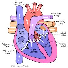
My favorite part was looking at the heart. The heart is so interesting to me and being able to hold it in my hands was so cool and fascinating. Looking at the heart I was able to differentiate some of the different parts.
IMG_1101
Holding the real pig heart I was able to feel the thickness of the aorta and the thinness of the atrium. We cut into the atrium slightly to see that it actually is a thin layer. Seeing characteristics like these help me understand more about heart and each of its structures and functions. To me the heart really was my favorite part and learning about the circulatory system. This system is one of my favorites to learn about.
Unknown-1
Overall, the fetal pig dissection was a successful activity to learn more about the body systems. I loved dissection the pig and it was a great way to have fun while learning!!
“Heart Diagram.” Wikimedia. N.p., n.d. Web. 28 Apr. 2015.
Pictures taken by me and other lab members

Thursday, March 26, 2015

How Life Begins on Earth - The Ancient Folktales

BigBangTheoryTitleCard
"Our whole universe was in a hot dense state,
Then nearly fourteen billion years ago expansion started. Wait..."

If you are a "The Big Bang Theory" fan, you will probably be very familiar with the lines above. If you are not, it is totally okay, because today we are not going to talk about the episodes of  "The Big Bang Theory" , we are going to talk about the real Big Bang Theory and reveal some interesting stories about how life begins on earth.

In truth, people in today's society will accept the big bang theory. At the very beginning, the early atmosphere was toxic, and a constant asteroid bombardment churned the landscape into a worldwide ocean of molten rock. As soon as the environment settled down to be relatively habitable, life appeared.
images
 
However, because of the lack of technically advanced tools, ancient people were unable to discover the scientific aspect of the formation of primate life, therefore, they created many myths of the formation of the universe.

In the Korean folktale, for instance, heaven and earth were stuck together but not bonded each other. Then a gap opened between the heaven and earth so that heavy subjects sunk, forming the earth. On another side, light subjects floated up, forming the sky. A blue drop of from the sky fell and caused a collision which made all creations of the universe except the Moon, the stars, and the Sun

Far from Asian countries, Egyptians had their own mythology too. They believed that at first, the world was a dark and milky place. One day, the God Atum cried and these tears gave birth to men and women. Later, Shu and Tefnut (Atum) had Nut (the sky) and Geb (the earth), who were lovers separated by Atum, but before they were separated, Nut and Geb gave birth to the main five Egyptian gods, and together made the earth a favorable place for human to live.

See! How interesting these myths are. What is your opinion of these myths? Do you know any myth created by other cultures? Please comment below!

Citation:
Citation of picture 1:
Picture 2:
Explosion. KeyWordPictures, 1 Jan. 2009. Web.http://www.keywordpictures.com/keyword/bigbang%20explosion

For further reading, please click key words below:

Sunday, February 15, 2015

Round-up: Darwin, Evolution and More!

This past unit has been a busy one for the AP Bio Team, as we covered chapters on Darwin, evolutionary theories, and even some math that goes along with the evolution of populations. We started off by covering the various theories of evolution and Darwin’s contributions that claimed organisms change and adapt, like the finches he researched. We watched various clips that discussed the Galapagos Islands and Darwin’s observations on the islands. There were also a couple of in-class discussions about these theories and Darwin’s journey around the world. These class discussions brought up a lot of good points about the way evolution was viewed has changed a lot over time and that Darwin’s journey was a pivotal change for how we view evolution. After various classes of watching videos and discussion, we moved on to the evolution of populations and the Hardy Weinberg equation.

Darwin's finches

We first discussed the ideas that populations have variations of alleles, or different versions of a gene, the frequency of these alleles can be determined and that allele frequencies change due to various factors such as natural selection, genetic drift and gene flow. Allele frequencies can be calculated using the Hardy Weinberg equation:
p2 + 2pq + q2 = 1
Initially this equation doesn’t look too inviting, however it is quite simple. “p2” stands for the frequency of homozygous dominant alleles in the population, “2pq” is the frequency of heterozygous alleles and you can probably guess that “q2” stands for the frequency of homozygous recessive alleles. This equation can be used to predict and describe what alleles and their frequency are in the population. 

Along with learning about the Hardy Weinberg equation, we discussed the five conditions that must be met for a population to stay the same and have no alterations in frequencies. These five conditions are:
Large population, random mating, no mutations, no movement, no natural selection

As you can probably assume, these conditions are not met in real life, however, it just shows the environment needed to have no changes in population. Also, as a class, we conducted a lab that involved the class “randomly mating,” using cards with “A” and “a” as different alleles, for five "generations," or rounds of mating, under various conditions. We counted how many people had each type of allele at the end and then calculated the before and after allele frequencies to practice using the Hardy Weinberg equation. It was a very fun and interactive lab that was enjoyed by everyone. 

After reviewing Hardy Weinberg, we then moved on to a virtual lab that looked at the evolution of Stickleback fishes. Each student went through the lab online, looking at how scientists identify differences in a Stickleback population by looking at the pelvic spines of living fish from various lakes and various fossils of Stickleback fish. These spines are present in some Stickleback populations, while other populations have very few individuals with a spine. This lab showed how the presence of the Stickleback spine in certain areas decreased over time due to their environment. Which means this lab demonstrated how an organism can adapt and change, or evolve, over time to suit the environment better. The Stickleback lab also introduced the idea of Chi-Square analysis, which is a statistical test done to determine the likelihood that the data is accurate and reliable. This statistical analysis is difficult to explain, but very useful in labs and experiments once understood.

Stickleback fish with and without pelvic spines 

Overall, this unit was very interesting, as it looked at the broader picture of life and evolution. The various in class activities and labs we did helped our understanding and I’m looking forward to see what we’re learning next! Thanks for reading!


Works Cited:
Gould, John. Darwin's Finches. Digital image. Wikimedia. Wikimedia, 27 Feb. 2012. Web. 15 Feb. 2015. <http://commons.wikimedia.org/wiki/File:Darwin%27s_finches_by_Gould.jpg>.

Shapiro. Convergent Pelvic Loss. Digital image. Shapiro Laboratory. University of Utah, n.d. Web. 15 Feb. 2015. <http://biologylabs.utah.edu/shapiro/Shapiro_Lab/Research.html>.


Tuesday, January 20, 2015

Gene Expression and Biotechnology Round-up!

After returning from a relaxing winter break, the AP Bio Team has jumped right back into the swing of things. We started a new unit, continuing our studies on gene regulation and focusing on DNA tools and biotechnology. Last week we completed multiple virtual labs and online explorations covering topics such as stem cells, cancer genetics and bacterial identification. It was really interesting to learn about the different types of stem cells and how they can lead to potential treatments to cure diseases like leukemia. At the same time, we uncovered many ethical questions regarding stem cell research. Many people do not feel comfortable with destroying a human embryo, and scientists have struggled with questions such as "When does a human life truly begin?" or "Can the destruction of one human embryo be justified if it saves a countless number of other patients?". It will be interesting to see where stem cell research goes in the future.
The main purpose of the bacterial identification lab was to familiarize us with the science and techniques used to identify different types of bacteria using their DNA sequence. The four basic steps involved in this process are:
1. Prepare a sample from a patient and isolate whole bacterial DNA
2. Make many copies of the desired piece of DNA
3. Sequence the DNA
4. Analyze the sequence and identify the bacteria
The lab showed us how the PCR (polymerase chain reaction) machines allow many copies of DNA to be made, and how gel electrophoresis is a method used to separate molecules based on difference of size or charge. We were able to identify the bacteria by determining whether the sequence we found had a significant degree of similarity to another known sequence on the BLAST database. 
Later in the week we conducted our own mini-lab using gel electrophoresis to identify whether or not each patient was normal, had sickle cell disease or was a carrier of sickle cell disease. The first sample was of normal hemoglobin, the second sample was sickled hemoglobin and the third sample was a carrier of sickle cell. Those three samples represented the control of the experiment. The fourth and fifth samples were patients with unknown hemoglobin. The wells are placed on the negative end of the gel electrophoresis box. Because the sickle cell hemoglobin is slightly more positive compared to normal hemoglobin, the sickle cell hemoglobin will travel slower across the gel. The carrier hemoglobin shows two bands instead of one. After the gel ran, the results showed that patient 1 was normal and patient 2 was a carrier. 
In another mini lab, we conducted a microarray analysis to a newly diagnosed breast cancer patient to determine her gene expression profile. We then proceeded to decide on her course of treatment. In order to perform the microarray analysis we had to collect healthy and cancerous tissue samples from the patient. That way we were able to look at what genes are turned on and off in the healthy cells compared to the cancerous cells. Based on the results we were able to match the patient with the cancer treatment that would be most effective. 
Next, we learned about epigenetics, the study of heritable traits that are not caused by changes in the DNA sequence. We watched the documentary "Ghost in Your Genes", which discussed how different experiences could affect gene expression. Scientists following the mapping of the human genome scientists discovered chemical markers on DNA that effectively turn genes on or off, dramatically influencing growth and development. The markers vary widely from person to person and are influenced not just by the environment but by experiences. Most remarkably the markers can be inherited generation after generation. A study was done on identical twins where researchers collected cells from twins ages 3 to 74. It revealed epigenetic tags that showed younger twins having extremely similar epigenomes, but the older twins had very different epigenomes. This supports the idea that experiences are linked to one's epigenome. A different study was done with rats, specifically looking at the behavior of the mothers. The research showed that the care the rats receive as babies can affect long term health conditions. A chemical mark somehow captures the memory of a nurturing mother. Stress levels of the neglected rats soared. This shows how personality traits could potentially be linked to one's epigenome. In the last study conducted in the documentary, information was gathered from a Swedish village that showed how a famine might affect people a century later, even if they haven't personally experienced it. According to the research, if the grandfather had experienced a surplus of food in early childhood, the chances of his grandson having diabetes would be four times greater. This study focused on the environment molding one's epigenome. These discoveries have changed the face of inheritance it can lead us to believe that the decisions we make today can have a big impact on the lives of our children and grandchildren. 
Continuing to study gene expression, the AP Bio Team acted out the functions of the lac operon!
The last major activity of these past 2 weeks was a really cool lab where we were able to make bacteria glow! The process of introducing foreign DNA into a host is called transformation. Biofluorescence is the ability of an organism to produce proteins that make the organism glow. The gene that codes for this Green Fluorescent Protein (GFP), known as the gfp gene, can be isolated from a source (an organism such as a jellyfish) and used to transform another organism. The bacterium E. coli is ideal for transformation because this bacterial cell is small and reproduces quickly. Arabinose is a sugar that bacteria break down for nutrients. The arabinose operon contains a promoter and three genes that code for three enzymes that digest arabinose. In the modified operon, these genes have been replaced by the gfp gene, so that when arabinose is present, the pathway to produce GFP will be triggered and the transformed bacteria will fluoresce.
Thanks so much for reading! It has been a crazy 2 weeks packed full of information. I can't wait to see what comes next!

Sunday, December 14, 2014

Roundup: Genetics and Review

Finals are next week (it’s crazy how fast the semester has gone!) and the AP Bio team has been working hard at finishing up our genetics unit and beginning review. We jumped right in after a relaxing Thanksgiving break with a class discussion about Cracking the Code of Life, a PBS documentary we watched about the progress we have made in genetic research. The movie talked a lot about the human genome project, which was an effort to map the entire human genome using new technology. Instead of talking only about the science of genetics, we got more into ethics and our opinions about DNA technology. We came up with some discussion questions to guide our talk, they were:
·     1) Is it possible/ethical that the U.S. would ever have a DNA database of citizens available to doctors or researchers?
·     2) Is it smart to genetically test people- should the president be tested? Do we want to and should we know if others or we have a disorder?
·     3) If parents could choose a child’s DNA, what would be the social consequences?

Each of us wrote blog posts about our thoughts on the film – here’s what Bharathi had to say on the prospect of parents choosing their child’s DNA:
"There has been a lot of controversy on this subject. I believe that parents should not be able to choose their baby’s DNA because it will make everyone perfect. The world is based on imperfections and imperfections are what create diversity and uniqueness in our lives." 
Abi shared her thoughts on the ethics of DNA technology, speaking about the medical records in Iceland, which were public to researchers:
“…There is an ethical issue with as to whether the family history from everyone was appropriate to release to the public. Personally, I think that if the DNA can help scientists figure out how genetic illnesses work, looking at an entire population’s DNA is acceptable.”
Finally, Shauna shared her opinion on whether she would like to know about possible genetic disorders:
"If there was a chance I could have a gene such as a cancer gene, I would want to know in order to do what I could to prevent it. I think it is better to know and be prepared than for it to just happen.” 
Whether we want to know all of this new genetic information or not, advancement in DNA technology is happening! This became clear later in the week, when we split up into groups and researched emerging DNA technology in fields such as medicine, pharmaceuticals, forensics, the environment, and agriculture. The presentations were very interesting – I had no idea of the breadth of subjects that DNA could be applied to, and I had no idea of the number of technological advancements that have been made recently. The information in our genome has the power to cure diseases, congeal copper, catch criminals, and even yummier cucumbers! One of the most interesting tidbits I learned was during the agriculture presentation, when Simran shared that researchers at UC Davis have found a way to genetically engineer and “domesticate” wild cucumbers. When each of 9 genes in the cucumber is turned off, the bitter taste characteristic of wild cucumbers disappears.


This past week, we focused on our genetics symposium, where individuals and small groups delved into various genetic disorders. We learned about the internal, external, psychological and biochemical characteristics of everything from Celiac disease (which affects about 1 in 100 people) to Progeria (which affects 1 in 3-5 million). Since genetics encompasses so many other topics that we have studied, our genetics symposium ended being a great review of things like cell signaling, cell structure, and meiosis/mitosis.  Both Amanda’s Voicethread (on Angelman’s Syndrome) and Rebecca’s Voicethread (on Hemophilia) are posted on their blogs, so check them out!


Thanks for reading and wish us luck on our final!

Picture from:
Gruepig. Pickling Cucumbers. Digital image. Wikimedia Commons. MediaWiki, 12 Nov. 2005. Web. 12 Dec. 2014.

Monday, December 1, 2014

2 Week Biology Sum-Up!

IMG_8489
Over the past two weeks, AP Biology has been action-packed. We have managed to squeeze in three chapters (give or take), an extra credit assignment. and an educational, outdoor fieldtrip!
fullsizerender-14The three chapters covered genetics, DNA replication, and protein synthesis. Genetics was more of a review of Mendel’s laws and punnett squares. I feel that genetics is the most interesting portion of biology because genetics and genes is what make our society diverse. DNA replication is interesting because in learning DNA replication my class and I could “connect the dots” about how DNA replication is the precursor for mitosis. In going in depth into DNA replication, I was able to grasp a better understanding of both concepts. Lastly, protein synthesis is where RNA is produced and used in order to code for specific proteins. This is important because through cell signaling, our bodies can produce the necessary proteins to keep the functionality of our bodies.
The extra credit assignment was to extract DNA from a strawberry using hose hold ingredients. I worked in a group of three with Bharathi Chinnakotla and Jennifer Parascandolo. The experiment is shown on all three of our blogs. Their links are: https://bharathisbioblog.wordpress.com/2014/11/26/strawberry-dna-extraction/ and https://biologybloggerr.wordpress.com/2014/12/01/strawberry-dna-extraction. This experiment really opened my eyes on how easy it is to extract DNA from a strawberry and that aspect reimg_85811ally interested me. As Jennifer stated, "Me and my partners screamed of happiness because we thought we were not going to be able to do the project." The joy we felt lifting the DNA up was that of no other. We felt smart and capable of doing such an intricate experiment. Bharathi states that the experiment could have been improved if we "placed the DNA under a microscope."
The outdoor field trip encompassed within this two week period, was at the bay front park in Palo Alto. This field trip, although educational, was very fun as well. My class and I collected data on many aspects of the soil found at the bay front that could be used in order to better the environment there and improve the lives of the species.
Works cited
All pictures taken by Jennifer
Parascandolo and myself.

Friday, November 21, 2014

Saving the Bay


On Wednesday November 19, 2014, the AP Bio Rockstars made their first trip to the Palo Alto Baylands to work with Save the Bay. Students collected soil data and calculated biodiversity using Simpson's Biodiversity Index. The data collected will be shared with Save the Bay and added to the data collected by classes and groups around the bay. 
Here's more about the experience!


Monday, November 17, 2014

Meiosis in Motion Roundup

In my AP Biology class, we learned about cell signaling, mitosis, and meiosis in the previous unit. We had a project on each so that we could understand the material better. For cell signaling, we had a project where we made a digital presentation on the effects of Ebola on the signaling pathway. For mitosis, the class split in half, and one group made a three dimensional diagram of mitosis in a plant cell and the other made a three dimensional diagram in an animal cell. For meiosis, we individually made a moving presentation of the process in an animal cell. This project was by far the most challenging but the most beneficial.

We were allowed to work individually or with a partner, and the goal was to create a project to watch a cell divide. After completing the project, each student embedded the video into a blog post, including a summary of meiosis and their thoughts on the project.

Summary of Meiosis

In case you do not know what meiosis is, Bharathi Chinnakotla gives a precise summary in her blog post! She says,
"Meiosis is a type of cell division that is crucial for sexual reproduction because it creates variation and genetic diversity. In this type of cell division, one diploid cell undergoes two divisions to create four haploid cells. The two divisions that take place in meiosis are meiosis 1 and meiosis 2."
"In prophase I, homologous chromosomes (a pair of two sets of sister chromatids that carry different versions of the same genetic information) pair up and exchange genetic information by “crossing-over.” This is a very important step in meiosis, as this step allows for increased genetic diversity of the offspring. The rest of meiosis continues very much like mitosis does: the rest of prophase I sees the dissolution of the nuclear envelope and the formation of spindle fibers, while metaphase I includes pairs homologous chromosomes lining up along the metaphase plate. During anaphase I, these homologous chromosomes are pulled apart from one another, and two distinct cells are formed through telophase and cytokinesis."
"In meiosis II, prophase II is the first step. In both cells the spindle forms and the chromosomes, made of two chromatids, move towards the center. In metaphase II the spindle fibers are lengthening and the chromosomes are in the middle at the metaphase plate. Next in anaphase II the spindle fibers begin to shorten and the chromatids move towards opposite poles in both cells. Lastly in telophase and cytokinesis II the two cells form haploid daughter cells leaving a total of four cells."
Amazing Project Videos

After summarizing Meiosis, each AP Bio Rockstar embedded her project into the post.


Comments and Thoughts

After posting the video, we made comments and shared our opinions on the project.
"For this project, the most challenging part was using Play Dough to create the phases of meiosis. The dough was often crmbly and difficult to mold, so some of the shapes we desired to create did not turn out as neat as we had hoped. However, because this project was incredibly hands-on, I think I was able to get a much better understanding of each phase of meiosis. Doing a project like this immerses me into a project far more than reading text from a book can. Overall, though this project was difficult, the challenge helped me understand and remember the process of meiosis much better."   
Elina Vanuska included a short reflection: 

"This project was very challenging because it required complete knowledge of what is occurring during meiosis and the skill and creativity to make a stop motion. The visual aspect of the project allows people to understand the topic easier, however this stop-motion was very difficult to create in the very short amount of time Nicole and I had to create it. However, with that being said, it was a good way to cover meiosis in a visual manner where we can actually see what is happening and I now have a better understanding of meiosis."

There are so many more amazing projects that I did not include in this blog post, so feel free to check out all the posts made by AP Bio Rockstars and see their masterpieces!

Friday, November 7, 2014

Mitosis and its phases!

This unit, in AP Biology, the class learned about cell communication and reproduction. The class worked hard and endlessly these past few weeks to understand the concepts of communication and reproduction.

One of the topics discussed in this unit was mitosis. The class learned about mitosis and the different phases of mitosis. The team also did a lab on mitosis and the duration of each of the phase. By using the class's knowledge about mitosis and its phases, the class had to come up with a way to figure out how long each phase of mitosis was by looking at onion root cells in a microscope. The AP Bio team had to put on their thinking caps to find solutions to problems.

The AP Bio team decided that the best way to figure out the duration of each phase of mitosis was by counting the total number of cells in view in the onion root slide and divide that number by the number of cells in each of the phases. Then, that number should be multiplied by 24 hours. That number would be the time each cells spends in each phase of mitosis. Another way people approached this problem was by finding the amount of time each phase takes in mitosis by using percentages (i.e. Entire Mitosis process would be 100%)

If you are following the AP Bio Blogs, you may have noticed that the class blogged about the mitosis phase lab. The class included a purpose, introduction, method and data.

Here’s how Shauna approached this lab:

1.  Take three samples of an onion root tip

2.  Take the first sample and focus it on the tip under the microscope

3.  Start by counting the total number of cells (an easy way to accomplish this is to count down and up and then multiply the two numbers to get an approximated value)

4.  Count the number of cells in each phase and record the number

5.  Divide the number of cells in each phase by the total number to get the percent of each one

6.  Next times the percent by 24 to find the total hours spent in each cell

7.  Repeat steps 2-6 with the other two samples
Check out Shauna’s Blog by clicking the link: http://journeythroughbiology.wordpress.com/

Here’s how Allie approached the lab:

1. Set up a slide of an onion root tip under a microscope with x.65 magnification.

2. Focus on a segment of the cell slightly above the tip of the root, and count the number of cells total in the segment.

3. Using the visual cues identified in the introduction, count the number of cells in each phase of the cell cycle (interphase, prophase, metaphase, anaphase, and telophase). Enter these numbers in the data table

4. Divide the number of cells in each stage by the total number of cells in the cell segment to determine the percent of cells that are in each phase of the cycle.

5. Repeat steps 1-4 two more times, using a different onion root tip cell slide each time.6. Calculate the average percent of time spent in each phase and multiply this number by 24 to get the hours spent in each phase of the cell cycle.

Check out Allie's Blog here:http://apbionce.wordpress.com/

The AP Bio team collected data that shows how long each phase of mitosis is:
Check out Rebecca’s data:
Data:
Anaphase: 0.309 hours (~18.54 min)
Interphase: 10.520 hours
Prophase: 5.768 hours
Metaphase 0.927 hours (~55.622 min)
Telophase: 3.091 hours
Time calculated with: 
(Total number of cells in a phase/ Total number of cells)  x 1440 minutes = total time of each phase                     
Data:
# of Cells
I
P
M
A
T
Total
Slide 1
61
33
5
3
27
135
Slide 2
68
23
4
2
3
99

I
P
M
A
T
Total
Average # of cells
64.5
28
4.5
1.5
15
116.5
Check out Rebecca's Blog: http://biobloggingbecca.wordpress.com/
Check out Simone’s data:


Interphase
Prophase
Metaphase
Anaphase
Telophase
Total
Trial 1
Trial 2
Trial 1
Trial 2
Trial 1
Trial 2
Trial 1
Trial 2
Trial 1
Trial 2
Trial 1
Trial 2
Number of cells
69
129
17
27
1
1
2
4
1
4
90
165
Percent of cells
77%
78%
19%
16%
1%
0.60%
2%
2.40%
1%
2.40%
100%

Check out Simone's Blog: http://simonesbioblahg.wordpress.com/

Overall, this lab was a lot of fun. At times it was difficult to identify the phases by looking at onion root cells in a microscope. However, by finding the phases in the cells, the class was able to understand what was happening in each of the phases. Also, figuring out how to find out the duration of each phase was challenging, but it made the class think and come up with creative solutions. This lab definitely helped the class better understand mitosis!